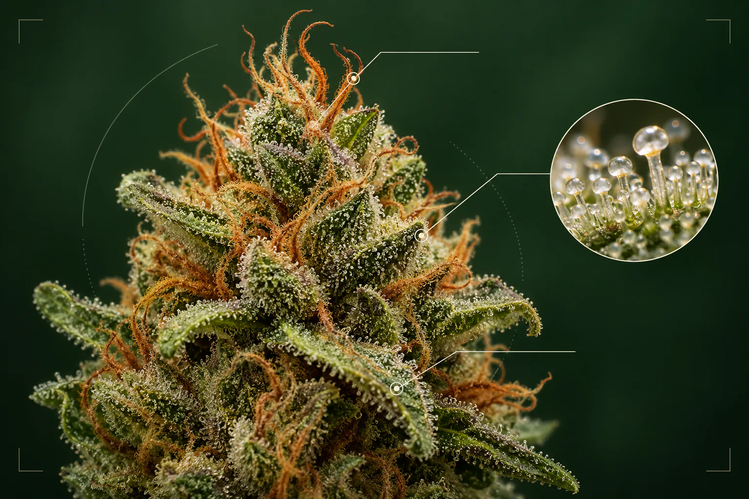
Look closely at a cannabis flower and the familiar green lump becomes a miniature landscape. Leaf tips curl between swollen structures. Fine orange threads cross the surface. Under magnification, translucent glands stand like tiny mushrooms.
Knowing the names of those parts helps you describe what a photo actually shows. It also prevents common identification mistakes: orange “hairs” are not trichomes, every frosty leaf is not a flower, and the structure widely called a calyx is usually more accurately described as a bract.
Cannabis flower anatomy at a glance
The dried product commonly called a bud is usually a female inflorescence—a cluster of many small flowers arranged on condensed branches. Its most visible parts include:
- Bracts: small leaf-like structures that surround individual female flowers and become densely coated in resin glands.
- Stigmas: the paired, hair-like tips that emerge to receive pollen; they often dry from pale cream to orange or brown.
- Pistil and ovary: reproductive structures enclosed inside the flower, mostly hidden in a dried bud.
- Sugar leaves: small leaves tucked into the flower cluster, often carrying a visible layer of trichomes.
- Trichomes: microscopic epidermal outgrowths; glandular types make and store much of the plant’s cannabinoids and terpenes.
- Stem and branches: the framework holding the clustered flowers together.
These structures overlap tightly after drying and trimming. A phone camera can record their color, shape, and surface texture, but it cannot see every hidden organ or chemically measure what the glands contain.
Bracts: the resinous building blocks of a bud
Many of the pointed, teardrop-shaped green structures covering a cannabis bud are bracts. In botanical terms, a bract is a modified leaf associated with a flower. Female cannabis flowers include a close-fitting perigonal bract around the ovary, while additional bracts and reduced leaves occur throughout the larger inflorescence.
Bracts matter because they carry abundant glandular trichomes. Peer-reviewed microscopy studies have examined how these glands develop across bract surfaces as flowers mature. When a flower is pollinated, the ovary inside develops a seed and the surrounding bract can swell.
In a dried sample, stacked bracts help create the bud’s surface pattern. Their size, spacing, and pointed tips may contribute to a loose, compact, round, or elongated look. Those traits can be useful for visual comparison, but they do not prove a strain name or potency.
Is it a cannabis calyx or a bract?
Cannabis vocabulary gets messy around the word calyx. Growers and retailers often use “calyx” for the resinous teardrop visible around a female flower. Botanically, that visible structure is generally the perigonal bract. The flower’s true perianth—the small floral envelope sometimes discussed as a calyx—is thin, reduced, and largely hidden.
This does not mean every casual use of “calyx” is incomprehensible. It means that “bract” is usually the clearer word when identifying the green, trichome-covered structure visible in a photograph. Scientific papers are not perfectly consistent either, so check how an author defines the term before comparing measurements.
Pistils, stigmas, and the orange hairs on weed
The threads commonly called pistils are more precisely stigmas, which are parts of the pistil. The pistil is the flower’s reproductive organ and includes the stigma, style, and ovary. In cannabis, two elongated stigma branches typically emerge from the surrounding bract.
Young stigmas are often pale or whitish. As flowers develop and then dry, they commonly curl inward and shift toward yellow, orange, red-brown, or tan. Genetics, handling, storage, and camera white balance can all influence how that color appears.
Stigmas can therefore provide a maturity clue in context, but they are not a potency gauge or a strain fingerprint. A bud with many orange threads is not automatically stronger, riper, or better. Our guide to orange hairs and cannabis pistils explains those limits in more detail.
Sugar leaves versus fan leaves
Sugar leaves are the small leaves that project from the flower cluster. Their nickname comes from the sparkling trichome layer that can make them look dusted with sugar. After harvest, a close trim removes much of this leaf material; a looser trim leaves more of it visible.
Fan leaves are the much larger, familiar multi-fingered leaves that power photosynthesis during growth. They are usually removed from finished flower. A dry bud with several narrow, resinous leaf tips is showing sugar leaves, not miniature fan leaves.
Sugar leaves can add useful visual information about trichome coverage, trim style, and handling. They can also make a bud look larger or frostier without revealing the chemistry of its bracts. That is one reason surface appearance cannot substitute for a batch-matched lab report.
Trichomes: the tiny glands behind the frost
Trichomes are small outgrowths on the plant’s surface. Cannabis has glandular and non-glandular types, but the glandular trichomes attract the most attention because they produce and accumulate cannabinoids and aromatic compounds.
Researchers commonly describe three glandular forms:
- Capitate-stalked trichomes have a stalk and a comparatively large rounded head. They become prominent on mature female flowers.
- Capitate-sessile trichomes have a rounded gland closer to the surface, with little visible stalk.
- Bulbous trichomes are much smaller and harder to resolve without microscopy.
To the naked eye, dense trichome coverage creates a frosty or crystalline surface. A macro photo may show individual heads, but normal lighting, focus, compression, and post-processing can exaggerate or hide them. Visible frost is biologically meaningful; it still cannot reveal an exact THC percentage. Read Cannabis Trichomes Explained for a closer guide.
Seeds, anthers, and other structures you may spot
An unpollinated female inflorescence usually directs growth toward flowers rather than mature seeds. If pollen reaches the stigmas and fertilization occurs, the ovule inside the ovary can develop into a seed. Mature seeds may split or push against the surrounding bract and become visible when a dried bud is opened.
Occasionally, a genetically female plant develops male reproductive structures. Mature anthers can release pollen, and their smooth yellow shape looks very different from paired hair-like stigmas. Research on cannabis hermaphroditism documents both altered flower morphology and resulting seed formation.
A photo may flag a visible seed, exposed anther, or unusual structure for closer inspection. It cannot show whether every hidden part of a dense flower cluster is seedless.
What cannabis flower anatomy can tell you in a photo
Once you know the parts, an image becomes more descriptive. You can look for:
- how tightly the bracts overlap and whether the structure is compact or airy;
- the amount of protruding sugar leaf and the apparent trim style;
- visible trichome coverage and whether macro detail preserves the gland heads;
- stigma color and distribution without treating it as a potency reading;
- seeds, crushed tissue, excess stem, discoloration, or possible foreign material;
- visual patterns that resemble reference examples in a strain-identification system.
Anatomy is not identity. Different cultivars can produce similar-looking flower, while environment, harvest timing, trimming, curing, storage, and photography can change the appearance of the same genetics. That is why an identify-weed-by-picture result should be treated as an estimate supported by labels, provenance, aroma, and laboratory data when available.
How to photograph flower anatomy clearly
Use soft, neutral light and steady focus. Photograph one intact bud against a plain background, then add a closer view rather than relying on one extreme crop. Avoid beauty filters, aggressive sharpening, colored grow lights, and direct flash glare. Include the flower’s overall shape as well as the surface detail.
For scale, place a clean ruler beside—not on—the sample. Do not handle the flower more than necessary, because pressure and friction can crush tissue or detach trichome heads. See how to take better weed photos for a complete scan-ready setup.
Put the visible clues together with KushScan
KushScan compares color, structure, pistils, trichomes, and other visible patterns in dry-flower photos. Use the result as identification context—not as proof of genetics, potency, or safety.
Download KushScanThe takeaway
A cannabis bud is a clustered female inflorescence, not one oversized flower. Resinous bracts surround individual flowers; paired stigmas emerge as hair-like threads; sugar leaves weave between the bracts; and glandular trichomes produce much of the chemistry associated with cannabis flower.
Learning that anatomy makes visual descriptions and photo comparisons more accurate. It does not remove the central limit of visual identification: shared features can narrow possibilities, but a picture alone cannot prove strain lineage, cannabinoid content, contaminant safety, or expected effects.
Sources
- Frontiers in Plant Science: Architecture and Florogenesis in Female Cannabis sativa Plants, microscopy-based description of female flower and inflorescence development, bracts, stigmas, and trichomes.
- Botanical Studies: Glandular Trichome Development, Morphology, and Maturation in Cannabis Inflorescences, research on gland types and their development on female bracts.
- Plants: Bracts, Buds, and Biases, peer-reviewed review of cannabis anatomy, trichome-density terminology, and cannabinoid comparisons.
- Frontiers in Plant Science: Hermaphroditism in Marijuana Inflorescences, study of flower morphology, stigmas, anthers, pollination, and seed formation.
- Oregon State University Extension: Hemp Anatomy, illustrated educational guide to Cannabis sativa vegetative and reproductive structures.